What You Need to Know About Age-Related Macular Degeneration
New York, NY— If you are age 50 or over, you may be among approximately 11 million in the United States who have age-related macular degeneration (AMD), a disease that affects the central part of the eye’s retina, or the part of the eye used when you are looking directly at a person or object. The condition is the leading cause of irreversible blindness in people over the age of 65 in this country, with an estimated 200,000 new cases each year, says Sherry Bass, OD, distinguished teaching professor at SUNY State College of Optometry specializing in ocular disease.
“Often early AMD is missed since some people may not have any symptoms at all,” explains Dr. Bass, who also serves as an optometric physician in the retina clinic of the University Eye Center.” But since the disease is progressive, early detection is essential to preserve vision.”
While early stages of AMD are often symptomless, disease progression can lead to:
- Blurry or fuzzy vision that is less clear and it may be hard to read fine print
- Straight lines may appear wavy or distorted
- Dark, blurry areas in the center of your vision
A patient with any form of AMD will maintain their peripheral vision or side vision since AMD affects the macula or central retina. Currently, there is no way to prevent AMD.
Who is at risk for AMD?
While age is the biggest risk factor, AMD causes are divided into genetic or hereditary components (those factors we cannot change) and non-genetic or environmental, lifestyle factors. Key genetic or hereditary factors include:
- Light-colored skin
- Light-colored or blue eyes
- Family history of AMD
Non-genetic or environmental factors include a history of smoking, high cholesterol, a high Body Mass Index or BMI (being overweight), cardiovascular disease and a diet poor in antioxidants. “Changing environmental factors does not necessarily prevent AMD since there may be genetic factors as well, but it is certainly a start,” adds Dr. Bass.
Types of AMD
There are two types of macular generation: “dry” and “wet.” What causes some people to develop the dry type while others progress to the wet type is not currently known, says Dr. Bass, but environmental factors may play a role.
About 90% of people with AMD have the atrophic or ‘dry’ type, which usually progresses slowly over many years. In dry AMD, small white or yellowish deposits (called drusen) form on the retina beneath the macula, causing the tissue to degenerate or atrophy over time. ‘Wet’ AMD starts out with drusen too, but the drusen lead to the growth of abnormal blood vessels that grow under the retina usually in the macular area. Because these new blood vessels are abnormal, they tend to bleed, and leak fluid, damaging the macula and causing it to separate from its base. The bleeding can result in scarring which leads to rapid, severe and irreversible loss of central vision.
“In untreated wet AMD, the vision can be as poor as counting fingers at 1 foot,” says Dr. Bass, “whereas in the dry form, the vision can typically deteriorate to 20/200 or 20/400, which is the size of the big “E” on the eye chart.” She adds that while a patient with AMD can be determined to be “legally blind,” which is vision of 20/200 or worse, a patient will never go completely blind from AMD in the dry or the wet form.
Check-ups and treatment
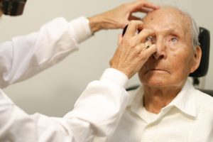
An optometrist will check for AMD by examining your retina through a dilated pupil and taking a picture your eye with an OCT (Optical Coherence Tomography). This device, used at the University Eye Center, can detect disease of the macula that may not be visible during the dilated retinal examination.
“If you have the dry type of AMD, your optometrist can monitor you and prescribe special magnifying glasses to help you read better. There is currently no FDA approved medical treatment to stop the progression of the dry form, although clinical trials are occurring. Patients diagnosed with wet AMD, however, will be referred to a retinal specialist,” says Dr. Bass. “Treatment for wet AMD consists of injections into the eye of a substance called an anti-VEGF agent, which stands for Anti-Vascular Endothelial Growth Factor. These drugs decrease the amount of VEGF in the eye resulting in less growth of new blood vessels from the choroid.” Recent developments in newer anti-VEGF agents have helped to preserve the vision of the close to 1 million patients across the US with this type of AMD.
For overall management of AMD, no matter dry or wet, Dr. Bass recommends a diet high in leafy green vegetables, like kale, spinach, and broccoli. She also suggests taking ocular vitamins that contain vitamin C, vitamin E, Zinc, Lutein, Zeaxanthin, and omega-3 fatty acids which have been shown to reduce the risk of progression in certain patients with the intermediate (not the early) form of AMD. It is important to remember that ocular vitamins and diet will not prevent the development of AMD since genetics and other environmental factors may be involved.
“The only way to diagnose and manage AMD is through routine eye examinations, especially after 50,” stresses Dr. Bass. “Don’t put it off even if you think you have perfect vision. Our eyes are a window to our overall health.”
For more information about care for AMD and other specialized services at the University Eye Center, visit sunyopt.edu or call 212-938-4001. Referrals are also accepted and welcomed.
February 16, 2020
Organization contact: Adrienne Stoller, communications@sunyopt.edu, 212-938-5600
###
About SUNY Optometry
Founded in 1971 and located in New York City, the State University of New York College of Optometry is a leader in education, research, and patient care, offering the Doctor of Optometry degree as well as MS and PhD degrees in vision science. The College conducts a robust program of basic, translational, and clinical research and has 65 affiliated clinical training sites as well as an on-site clinic, the University Eye Center. SUNY Optometry is regionally accredited by the Commission on Higher Education of the Middle States Association of Colleges and Secondary Schools; its four-year professional degree program and residency programs are accredited by the Accreditation Council on Optometric Education of the American Optometric Association. All classrooms, research facilities and the University Eye Center, which is one of the largest optometric outpatient facilities in the nation, are located on 42nd Street in midtown Manhattan. To learn more about SUNY Optometry, visit www.sunyopt.edu.